Free Webex Call
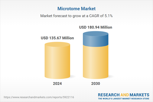
Global Microtome Market has valued at USD 417.86 million in 2022 and is anticipated to project robust growth in the forecast period with a CAGR of 5.06%through 2028. The Global Microtome Market is a dynamic and rapidly evolving sector within the broader life sciences and medical equipment industry. Microtomes are precision instruments used for the sectioning of biological specimens into thin slices for microscopic analysis. These devices play a crucial role in various fields, including histology, pathology, and research, aiding in the diagnosis and study of diseases, as well as in pharmaceutical and biomedical research. Speak directly to the analyst to clarify any post sales queries you may have.
10% Free customizationThis report comes with 10% free customization, enabling you to add data that meets your specific business needs.
The market for microtomes has witnessed significant growth and innovation in recent years, driven by advances in healthcare and research. Key factors influencing this growth include an aging global population, rising prevalence of chronic diseases, and the continuous expansion of medical and scientific research. In addition, the demand for high-quality tissue samples for accurate diagnosis and research has fueled the need for advanced microtome technologies.
Market participants in the Global Microtome Market encompass a wide range of companies, including established players and emerging startups, offering a variety of microtome models, from manual to fully automated systems. These systems differ in terms of precision, speed, and user-friendliness, catering to the diverse needs of laboratories and research facilities.
The market is also witnessing technological advancements, such as digital microtomes that provide enhanced precision and image analysis capabilities. Moreover, the integration of artificial intelligence and machine learning for data analysis is becoming a trend in the industry, aiding researchers and pathologists in their work.
Key Market Drivers
Advancements in Healthcare and Research
In the field of healthcare, precision is paramount. As medical science advances, there's a growing need for precise diagnostic tools to identify diseases and disorders at an early stage. Microtomes play a crucial role in preparing thin tissue sections for histopathological examination, enabling pathologists to make accurate diagnoses. These precise diagnoses, in turn, facilitate the development of tailored treatment plans, personalized medicine, and more effective therapies.The realm of biomedical research has undergone significant evolution. Researchers, pharmaceutical companies, and academic institutions are continually pushing the boundaries of science to develop innovative treatments and therapies. Microtomes are indispensable in this research process, helping scientists prepare tissue samples for a wide range of studies, including cancer research, drug development, and genetic studies. Their precision and reliability are invaluable in producing consistent and reproducible results.
Advancements in cancer research and genomics have driven the microtome market. With a deeper understanding of the genetic and molecular basis of cancers, researchers require highly accurate tools to prepare tissue samples for genetic testing and analysis. Microtomes provide the precision necessary to isolate specific cells and structures, aiding in the identification of genetic mutations and targeted therapies.
The study of the brain and neurological diseases has seen remarkable progress in recent years. Neuroscientists rely on microtomes to prepare ultra-thin brain sections for detailed analysis. These sections are critical for examining neuronal structures and identifying abnormalities associated with conditions like Alzheimer's disease, Parkinson's disease, and various neurological disorders.
Technological Advancements
These instruments offer unparalleled precision and consistency in tissue sectioning, reducing the margin for human error and significantly enhancing the speed and efficiency of the process. Automated microtomes are in high demand, particularly in high-throughput laboratories and research facilities.The integration of digital technology into microtome systems has been transformative. Digital microtomes come equipped with imaging sensors and software that capture high-resolution images of the tissue sections as they are cut. This not only provides an instant visual record of the samples but also allows for 3D reconstruction and digital analysis, making it easier to identify anomalies, perform quantitative assessments, and share results with colleagues and collaborators.
Technological innovations have led to the development of highly specialized blade materials, such as diamond and tungsten carbide, which ensure precise and consistent sectioning. These materials have extended blade lifespan, reduced the need for frequent blade changes, and enhanced the quality of the sections produced.
AI and ML technologies have found their way into the microtome market, particularly in digital microtomes. These systems can employ AI algorithms to optimize cutting parameters, recognize and differentiate tissue structures, and even provide automated feedback for blade maintenance and replacement. Such capabilities streamline the tissue sectioning process, reduce human intervention, and improve the overall efficiency of the microtome.
With the advent of the Internet of Things (IoT), microtome manufacturers are offering remote monitoring and diagnostics capabilities. This allows users to track the performance of their microtomes in real-time, receive maintenance alerts, and access remote support services, thereby reducing downtime and ensuring optimal instrument performance.
Technological advancements have led to the development of microtomes with ergonomic designs and user-friendly interfaces. These improvements enhance the user experience, reduce operator fatigue, and make the instruments more accessible to a broader range of users.
Aging Population and Chronic Diseases
With a significant portion of the world's population reaching an older age, there's a natural increase in the incidence of age-related diseases. The elderly are more susceptible to conditions like cancer, neurodegenerative disorders, and cardiovascular diseases, which require thorough pathological examination for accurate diagnosis and treatment. Microtomes play a pivotal role in preparing tissue samples that enable pathologists to identify the specific characteristics and stages of these diseases.Cancer is a leading cause of morbidity and mortality worldwide, and its prevalence increases with age. Microtomes are essential for preparing tissue sections for histopathological analysis, which is crucial for diagnosing cancer, determining its stage, and planning appropriate treatment strategies. With cancer cases on the rise, the demand for microtomes in cancer diagnostics is consistently high.
Conditions like Alzheimer's disease and Parkinson's disease are more common in older individuals. Researchers studying these neurodegenerative disorders rely on microtomes to cut thin brain sections for microscopic analysis. These precise sections help in identifying cellular and structural changes associated with these conditions, aiding in both diagnosis and research for potential therapies.
Cardiovascular diseases, including heart disease and stroke, are significant contributors to global mortality, and their risk increases with age. Microtomes are used to prepare heart and vascular tissue samples for detailed examination. These sections assist in understanding the underlying causes, the extent of damage, and the development of therapeutic interventions.
As the aging population experiences an increased need for organ transplants and replacements, microtomes are used to prepare scaffold structures from donor tissues. These scaffolds serve as a foundation for tissue engineering, regenerative medicine, and the creation of artificial organs for transplantation, reducing the demand for organ donors.
Researchers are continually exploring the biology of aging and age-related conditions. Microtomes are crucial in providing the precision needed to study the effects of aging on tissues and cells, enabling the development of new interventions and therapies to improve the quality of life for the elderly.
Key Market Challenges
High Initial Costs
Smaller research facilities, laboratories, and academic institutions often operate with tight budgets. The substantial upfront investment required to acquire a microtome, especially an automated or digital system, can strain their financial resources. In some cases, these budget constraints may lead to the postponement or cancellation of equipment purchases, hindering the market's growth and impeding progress in scientific and medical research.In healthcare institutions, managing limited budgets poses a constant challenge. The high cost of acquiring a microtome can divert funds from other essential areas, such as personnel, patient care, or acquiring additional medical equipment. This allocation dilemma often forces healthcare organizations to make difficult decisions regarding where to invest their resources.
In emerging economies, where healthcare infrastructure is still developing and resources are limited, the high cost of microtomes can be a significant deterrent. As these regions increasingly invest in healthcare and research, there's a growing demand for affordable and efficient equipment. The prohibitive initial cost can slow down the adoption of microtome technology, impeding the market's expansion in these promising areas.
In many established facilities, older and less expensive manual microtomes are still in use. These institutions may be reluctant to invest in newer, automated models due to the substantial financial commitment required. This reluctance to upgrade can hinder the adoption of advanced microtome technology and slow the overall market growth.
Competition with Alternative Technologies
Digital pathology scanners are gaining popularity as an alternative to traditional microtomes. These devices allow for the digitization of whole slides, enabling pathologists and researchers to view and analyze tissue samples electronically. This digital approach offers numerous advantages, such as rapid image acquisition, the ability to store and share digital slides, and remote access for consultation. Consequently, it reduces the reliance on microtomes for producing physical tissue sections.Three-dimensional imaging systems are advancing rapidly and are particularly appealing in research contexts. These systems allow for the reconstruction of 3D images from serial sections, eliminating the need for traditional microtome-based sectioning. Researchers can create detailed 3D representations of biological specimens, which is especially beneficial in fields like neuroscience, where precise three-dimensional structures are essential.
Traditional microtomes require the physical slicing of tissue samples, which can be time-consuming, and the process is inherently destructive. Alternative technologies offer non-destructive imaging and faster analysis. This advantage is particularly valuable in applications where speed and preserving the integrity of the specimen are critical.
Many of these alternative technologies are equipped with artificial intelligence (AI) and machine learning capabilities. These AI algorithms can automatically identify and classify features in tissue samples, reducing the need for manual labor and specialized expertise. This automated analysis is particularly appealing for high-throughput settings.
Key Market Trends
Advancements in Digital Pathology
Advancements in digital pathology have emerged as a driving force behind the growth of the global microtome market. Digital pathology, a transformative trend in healthcare and research, relies heavily on the critical role that microtomes play in producing high-quality tissue sections for imaging and analysis. This integration of digital technology with pathology has reshaped the industry in several key ways, boosting the demand for microtome systems.digital pathology systems have streamlined the process of capturing and analyzing tissue sections. Microtomes, as precision instruments for producing these sections, are pivotal in this workflow. As whole slides are digitized, pathologists and researchers can access high-resolution images of the samples in a digital format. This transition from traditional glass slides to digital images not only saves time but also enables remote access, thereby transforming the way pathologists and researchers operate.
Additionally, digital pathology systems allow for the storage and easy sharing of digital slides among experts and colleagues. The collaborative aspect of these systems facilitates consultations and discussions among professionals, even when they are geographically distant. The convenience of this digital approach not only expedites the decision-making process but also improves patient care through faster diagnoses and treatment planning.
Digital microtomes, a significant component of this trend, capture images of tissue sections in real-time as they are cut. This has opened up a realm of possibilities for in-depth image analysis. Researchers and pathologists can reconstruct three-dimensional representations of tissue structures and use digital tools for quantification, data sharing, and streamlined workflows.
Tissue Engineering and Regenerative Medicine
Tissue engineering and regenerative medicine have emerged as dynamic fields at the forefront of medical research and innovation, and they play a pivotal role in boosting the global microtome market. Microtomes, precision instruments used for the sectioning of biological specimens, are indispensable tools in the creation of engineered tissues and regenerative medicine applications.In tissue engineering, microtomes are used to prepare scaffold structures from donor tissues or biomaterials. These scaffolds serve as the foundation upon which cells, including stem cells, are seeded or cultured. Microtomes enable precise and consistent sectioning of these scaffolds, ensuring the creation of three-dimensional structures that mimic the native tissues of the human body. These engineered tissues have a wide range of applications, from replacing damaged or diseased organs to developing in vitro models for drug testing and disease research.
In regenerative medicine, microtomes are fundamental in the production of tissue sections that are used for transplantation, grafting, or other regenerative treatments. This field focuses on harnessing the body's natural healing abilities and leveraging tissue engineering principles to repair or replace damaged or degenerated tissues. Microtomes contribute to the development of viable tissue grafts with precise thickness, which is essential for successful transplantation procedures.
The role of microtomes in tissue engineering and regenerative medicine extends beyond the initial preparation of scaffold structures. Researchers and clinicians working in these fields rely on microtome-produced tissue sections to assess the quality and integration of the engineered tissues. Microtome sections are instrumental in analyzing cellular alignment, tissue architecture, and the distribution of various components within the engineered constructs.
Segmental Insights
Product Insights
Based on the Product, Microtome devices emerged as the dominant segment in the global market for Global Microtome Market in 2022. Microtome devices are the primary equipment used for the precision sectioning of biological specimens. They are fundamental to the workflow of pathologists, researchers, and scientists in various fields. Microtome devices are essential for obtaining thin and consistent tissue sections, which are critical for a wide range of applications in healthcare, research, and pathology.Microtome devices are versatile and find applications in multiple domains. They are used in histology labs for routine diagnostic pathology, in research labs for various scientific investigations, and in fields like tissue engineering and regenerative medicine. The wide range of applications ensures that the demand for microtome devices remains consistently high..
Technology Insights
Based on the Technology, the Fully Automated segment emerged as the dominant player in the global market for Global Microtome Market in 2022. Fully automated microtomes offer a level of precision and consistency that is challenging to achieve with manual or semi-automated systems. The automated cutting process ensures that each tissue section produced is of uniform thickness and quality, reducing the risk of human error. This level of precision is particularly critical in diagnostic pathology and research, where accurate results are paramount. Fully automated microtomes significantly enhance the efficiency and throughput of tissue sectioning. These systems can process a higher volume of specimens in a shorter time frame compared to manual or semi-automated counterparts. This speed and efficiency are especially advantageous in high-throughput laboratory settings, where a large number of specimens need to be processed quickly.Regional Insights
North America emerged as the dominant player in the global Microtome Market in 2022, holding the largest market share. North America boasts one of the most advanced and sophisticated healthcare infrastructures globally. The region is home to cutting-edge medical facilities, research institutions, and diagnostic laboratories that heavily rely on microtomes for accurate tissue sectioning in areas like pathology, histology, and research. The presence of such facilities drives a substantial demand for microtome technology. North America has a relatively high prevalence of chronic diseases, such as cancer, cardiovascular diseases, and neurodegenerative disorders. Accurate diagnosis and research in these areas are crucial for patient care and public health. Microtomes play a vital role in preparing tissue samples for histopathological analysis, aiding in early detection and disease characterization.Report Scope:
In this report, the Global Microtome Market has been segmented into the following categories, in addition to the industry trends which have also been detailed below:Global Microtome Market, By Product:
- Microtome Devices
- Accessories
Global Microtome Market, By Technology:
- Fully Automated
- Semi-Automated
- Manual
Global Microtome Market, By Region:
- North America
- United States
- Canada
- Mexico
- Europe
- France
- United Kingdom
- Italy
- Germany
- Spain
- Asia-Pacific
- China
- India
- Japan
- Australia
- South Korea
- South America
- Brazil
- Argentina
- Colombia
- Middle East & Africa
- South Africa
- Saudi Arabia
- UAE
- Kuwait
- Turkey
- Egypt
Competitive Landscape
Company Profiles: Detailed analysis of the major companies present in the Global Microtome Market.Available Customizations:
Global Microtome Market report with the given market data, the publisher offers customizations according to a company's specific needs.This product will be delivered within 1-3 business days.
Table of Contents
1. Product Overview
2. Research Methodology
3. Executive Summary
4. Global Microtome Market Outlook
5. Asia Pacific Microtome Market Outlook
6. Europe Microtome Market Outlook
7. North America Microtome Market Outlook
8. South America Microtome Market Outlook
9. Middle East and Africa Microtome Market Outlook
10. Market Dynamics
11. Market Trends & Developments
13. Porter’s Five Forces Analysis
14. Competitive Landscape
Companies Mentioned (Partial List)
A selection of companies mentioned in this report includes, but is not limited to:
- Diapath S.P.A.
- Leica Biosystems Nussloch GmbH
- Sakura Finetek Europe B.V.
- MEDITE GmbH
- SLEE medical GmbH
- Boeckeler Instruments
- Thermo Fisher Scientific Inc.
- S.M. Scientific Instruments Pvt. Ltd.
- AGD Biomedicals
- Amos Scientific Pvt. Ltd.
Table Information
Report Attribute | Details |
---|---|
No. of Pages | 185 |
Published | November 2023 |
Forecast Period | 2022 - 2028 |
Estimated Market Value ( USD | $ 417.86 Billion |
Forecasted Market Value ( USD | $ 557.18 Billion |
Compound Annual Growth Rate | 5.0% |
Regions Covered | Global |